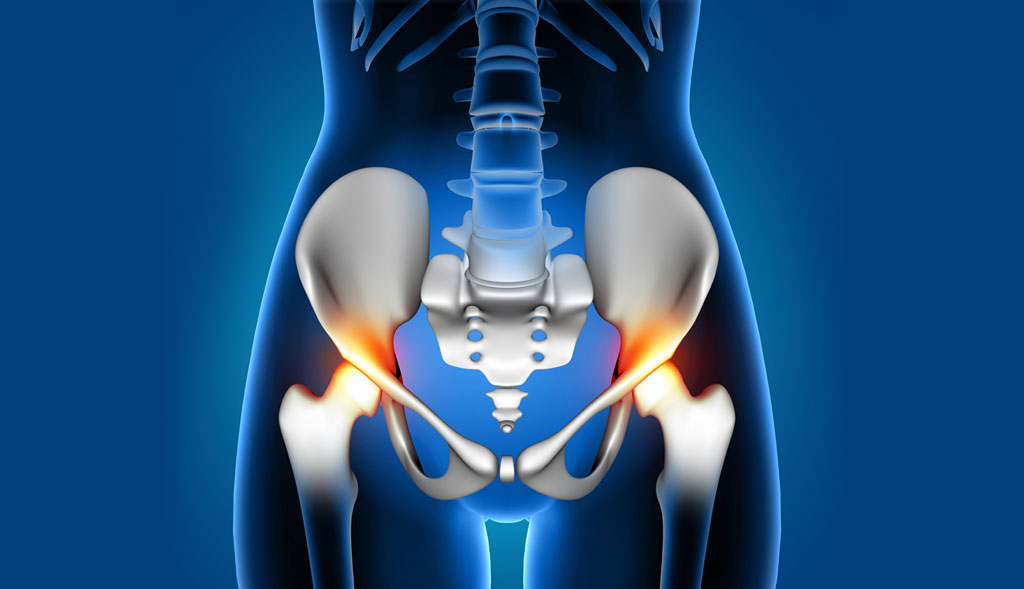
Pelvic disorders consist of a wide range of conditions, affecting the organs and structures in the pelvic region. The disorders can significantly affect a person’s life, causing symptoms like chronic pain, urinary and bowel dysfunction and sexual health issues. Accurate diagnosis is vital for effective treatment. Advancements in medical imaging have provided new tools to aid in this process. One such advancement is the open non-claustrophobic standing MRI, a technology that offers unique benefits in diagnosing pelvic disorders.
Challenges of Diagnosing Pelvic Disorders
Diagnosing pelvic disorders can be a challenging task, owing to the intricate anatomy and the variety of potential conditions. Traditional diagnostic methods, including pelvic exams and ultrasounds provide valuable information. Pelvic ultrasounds are first line of tests. MRI offers high-resolution images with better tissue characterization. However, it can be uncomfortable for some patients due to enclosed space, potentially leading to motion artifacts and incomplete studies.
Open Non-Claustrophobic Standing MRIs
The advantage of open non-claustrophobic standing MRIs is that this system addresses many of the limitations of traditional MRIs. These systems are designed with patient comfort in mind, offering a spacious and open configuration that reduces anxiety and claustrophobia. Furthermore, the ability to perform scans while the patient is standing or in various positions provides a more functional and dynamic assessment, which is particularly beneficial for diagnosing pelvic disorders.
Enhanced Patient Comfort: One of the primary advantages of open MRI systems is their ability to accommodate claustrophobic patients. The open design and the possibility of a standing or sitting position can significantly reduce patient anxiety, leading to a more cooperative experience and better quality images.
Accurate Diagnosis of Pelvic Organ Prolapse: Pelvic Organ Prolapse (POP) is a condition where pelvic organs descend into or outside the vaginal canal, due to weakened support structures. Symptoms can vary significantly based on the patient’s position. A standing MRI can more accurately depict the degree of organ descent and provide a clear picture of the condition, compared to traditional supine MRI or ultrasound, leading to more precise surgical planning and treatment.
Functional Imaging: Pelvic disorders can often be related to postural issues or conditions that are exacerbated in certain positions. Traditional supine MRI scans miss these dynamic aspects. The standing MRI allows for imaging in a weight-bearing position, providing insights into how gravity and posture affect the pelvic organs and structures.
Assessment of Musculoskeletal Disorders: Conditions such as pelvic girdle pain, sacroiliac joint dysfunction and pelvic floor muscle disorders benefit from the functional assessment capabilities of standing MRI. By visualizing the pelvis under normal load-bearing conditions, clinicians can better understand the relationship between symptoms and structural abnormalities.
Comprehensive Evaluation of Pelvic Pain: Chronic pelvic pain can arise from multiple sources, including muscles, nerves and organs. The open MRI offers a comprehensive evaluation of the pelvis, including, muscles, ligaments and surrounding structures, in a single, non-invasive study. This holistic view helps in identifying the cause of the pain, whether it is musculoskeletal, organ-related or a combination of factors.
Eastern Diagnostics at the Forefront
Eastern Diagnostics is committed to implementing the latest advancements in medical imaging to provide patients the highest standard of care. For patients suffering from pelvic disorders, open non-claustrophobic standing MRI offers a promising option for obtaining detailed and comprehensive imaging necessary for proper diagnosis and treatment.