Tilting MRI
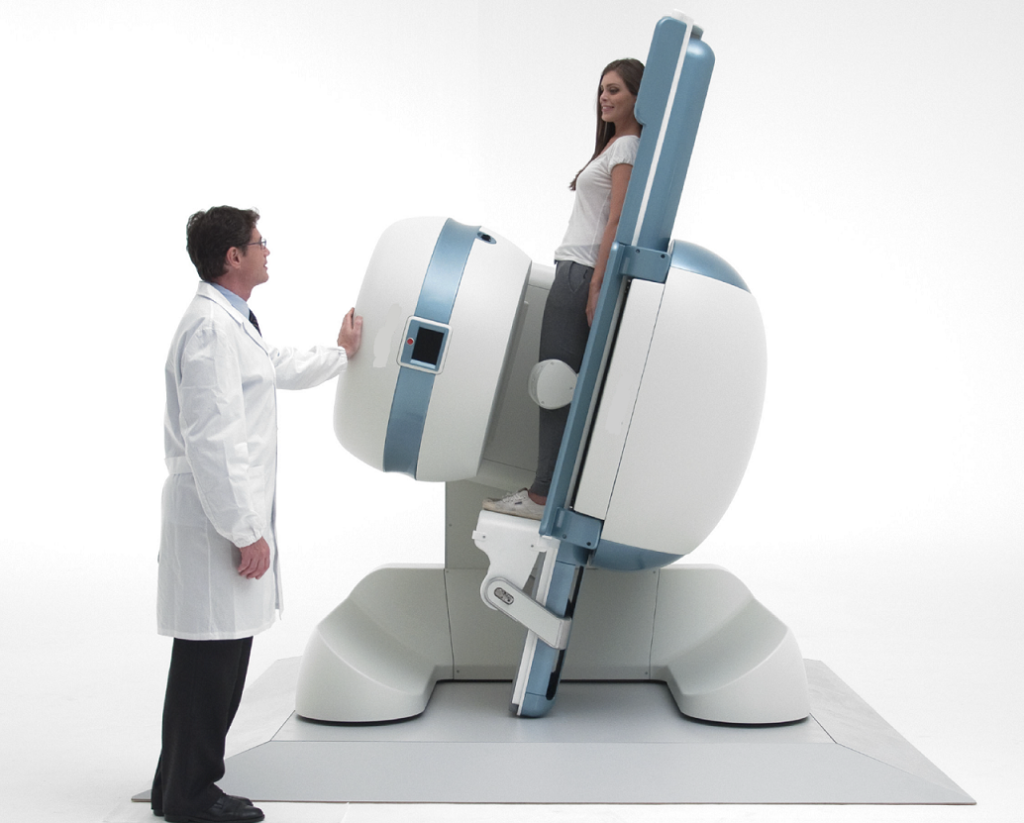
A tilting MRI, often referred to as a positional MRI or positional imaging system, features a patient table that can be dynamically moved and tilted while conducting the scanning procedure. This remarkable capability presents a multitude of advantages when compared to conventional static MRI machines:
Enhanced Visualization:
The ability to tilt the patient table allows for imaging from various angles, providing a more comprehensive view of anatomical structures. This can help medical professionals gain a better understanding of complex anatomical relationships.
Functional Studies:
Tilting MRI enables functional imaging by capturing images of the body in different positions. This is particularly useful for studying joint mobility, spinal alignment, and other dynamic processes that are not adequately assessed with static scans.
Reduced Artifacts:
Motion artifacts can distort MRI images, affecting diagnostic accuracy. Tilting MRIs can mitigate these artifacts by optimizing patient positioning, resulting in clearer and more accurate images.
Improved Patient Comfort:
Some patients, such as those with claustrophobia or mobility issues, may find traditional MRI machines uncomfortable. Tilting MRIs can offer a more comfortable experience, as patients have the option to be positioned in a way that minimizes discomfort.
Silent MRI
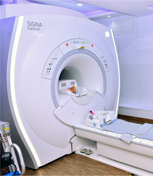
Magnetic Resonance Imaging (MRI) is a technology used in medicine to form images of various parts of the human body. The machine, with the help of magnetic fields, creates detailed images of the human body that help the doctor diagnose the problem.
Patient Comfort
An MRI can be an uncomfortable experience for many patients. At Eastern Diagnostics, we have taken utmost care to ensure a high-level degree of comfort for the patient.
Silent MRI
MRI machines typically produce a lot of noise, almost as much as a small construction site. This can be uncomfortable for the patient and it can cause them to be restless which would result in an improper scan.
Eastern Diagnostics employs a Silent MRI that suppresses the noise and brings it in line with a normal electrical device.
Visually Calming
The sight of a large machine can be imposing and can result in patient anxiety. Therefore to put the patient at rest, Eastern Diagnostics’ MRI room has been designed so as to be visually soothing. The gentle colours, soft lighting and nature-themed wall murals, immediately put the patient at ease, thus resulting in an overall positive experience for the patient.
Feet-first entry
Most MRI machines require the patient to be inserted head-first into the tunnel. This means that when the patient is fully inserted into the machine, his head is farthest away from the opening of the tunnel. This can create a sense of helplessness.
At Eastern Diagnostics the patient is inserted feet-first into the tunnel thereby ensuring that the head is almost at the opening. This means that the patient has a good view of the room outside the machine, thereby putting him at ease.
Other Facilities In Our Imaging Department
In addition to MRI, we also provide the below tests and diagnostics in our Imaging department.
- Low Radiation CT Scan
- 4D Ultrasonography with Elastography
- Bone Densitometry / Dexascan
- CT/US Guided FNAC
- Mammography